Predesigned silica nanofractal substrates are utilized for rapid cell patterning, based on differential cell adhesion originating from surface topographic interactions. Cell patterns with various shapes are successfully formed, from simple geometrical shapes to a complex “CELL” symbol. This study assists understanding of cell–substrate interactions and facilitates biological applications. Small, 2015 
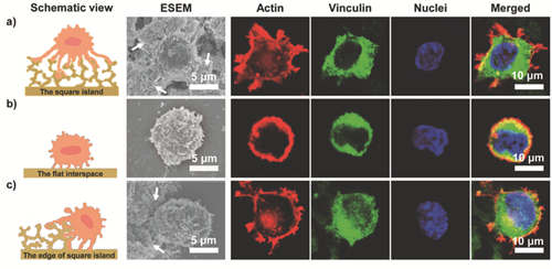
ESEM images and immunofluorescence images of NIH/3T3 cells adhered on different regions of the patterned substrate, Patten-(96, 29), including a) the square island of silica nanofractals, b) the edge of the square island of silica nanofractals, c) the flat interspace of the adjacent square islands. Immunofluorescent images include actin (red, for actin cytoskeleton), vinculin (green, for focal adhesions) and cell nuclei (blue). NIH/3T3 cells adhering onto the square island of silica nanofractals exhibited more filopodia (indicated by white arrows), more extended actin and a larger area of focal adhesion (a) than that on the flat interspace (b). The above phenomenon could be proved by the cell adhering onto the edge of the square island (c): one part of the cell adhering onto the silica nanofractals displayed the strong adhesive state as in (a), while the other part adhered to the flat interspace and showed the low adhesive state as in (b). The results indicate that within a short time, cells prefer to adhere onto the silica nanofractals and not the flat interspaces. Magnifications are the same in immunefluorescence images. |

