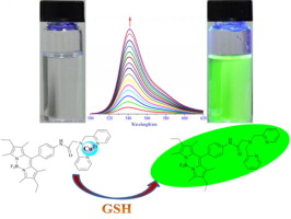
A new BODIPY derivative acting as “on–off” fluorescent sensor for Cu2+ in living cells and its·Cu2+ complex can also recognize biothiol.
A new fluorescent boron dipyrromethene (Bodipy) derivative 1 containing a di(2-picolyl)amine group as a binding site for Cu2+ was synthesized and characterized. Compound 1 behaves as an “on-off” fluorescent sensor for highly selective and sensitive detection of Cu2+ ions. The selective interaction between compound 1 and Cu2+ leads to formation of a 1·Cu2+ complex, associated with quenching of the fluorescence of 1. An investigation into the detection of biothiols shows that the 1·Cu2+ complex has better recognition for glutathione than cysteine and homocysteine in CH3 OH-HEPES (1:1, v/v) buffer solution at pH 7.4 (HEPES = 4-(2-hydroxyethyl)-1-piperazineethanesulfonic acid). In addition, fluorescence images obtained via confocal microscopy reveal that compound 1 exhibits excellent selectivity and high sensitivity for Cu2+ under physiological conditions and in living cells.
Journal of Photochemistry and Photobiology A: Chemistry
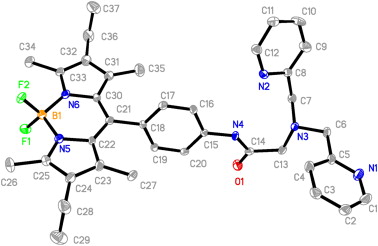
Perspective view of compound 1 with atom-labeling scheme. Hydrogen atoms and solvent molecules are omitted for clarity.
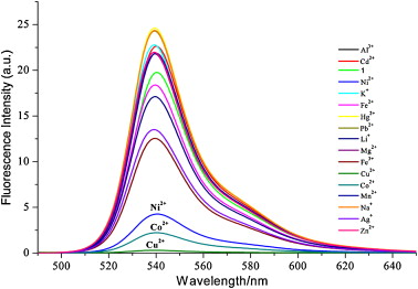
Fluorescence spectra of compound 1 (5.0 × 10⁻⁶ M) with addition of various metal ions in 10 mM of CH3OH-HEPES (1:1, v/v) buffer solution at pH 7.4. (λex = 526 nm).
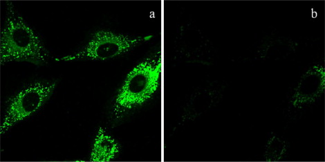
Fluorescence image of compound 1 in living cells at 37 °C. (a) A549 cells incubated with 5.0 μM of compound1 in 10−3 mM, 1 mL PBS for 30 min. (b) A549 cells incubated with 5.0 μM of compound 1 treated with 25 μM of Cu2+ aqueous solution with each of 125 μM Na+, Mg2+, K+, for 10 min.
Co-staining fluorescence images of compound 1 with LysoTracker or MitoTracker in living cells at 37 °C: (top) (a) The overlay of a fluorescence image of compound 1 with LysoTracker; fluorescence images of compound1 (b) and LysoTracker (c). (bottom) (a) An overlay of a fluorescence image of compound 1 with MitoTracker; the fluorescence images of MitoTracker (b) and compound 1 (c).